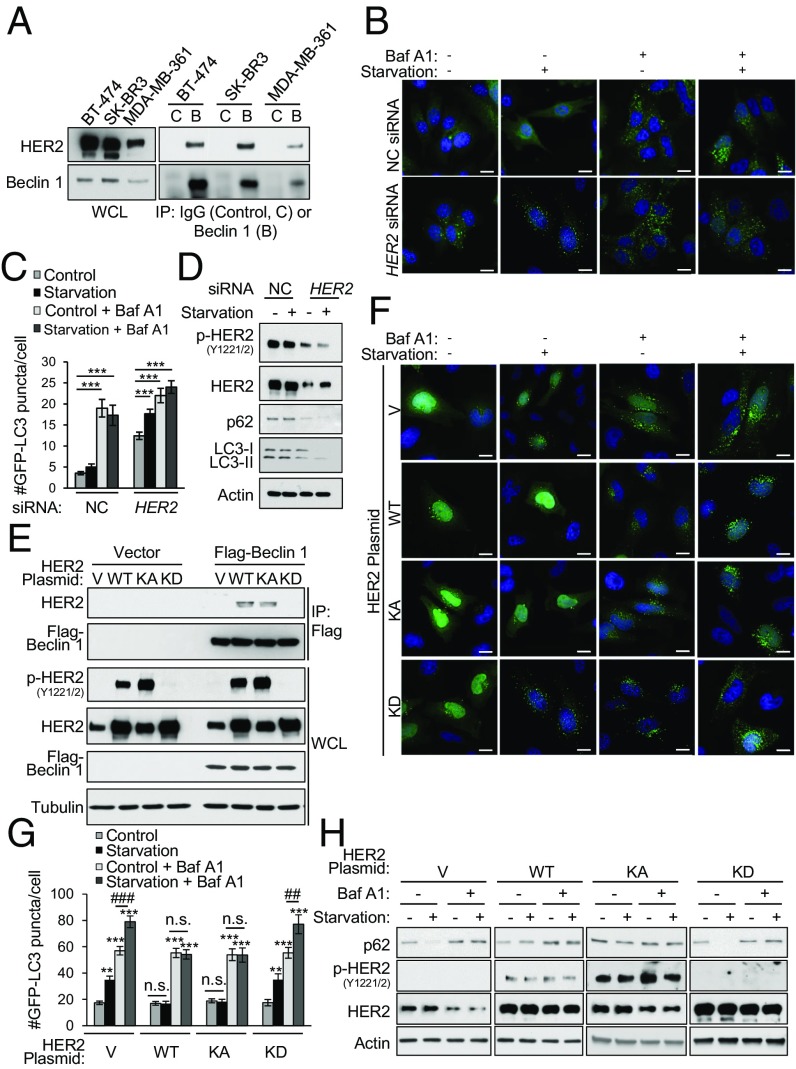
Fig. 1.
HER2 interacts with Beclin 1 and reduces starvation-induced autophagy. (A) Coimmunoprecipitation of endogenous HER2 with endogenous Beclin 1 in indicated HER2-positive breast cancer cell lines. B, Beclin 1 IgG; C, Control IgG; IP, immunoprecipitation; WCL, whole-cell lysates. (B and C) HER2 knockdown effects on autophagy in BT-474 cells cotransfected with GFP-LC3 and a nontargeting control (NC) or HER2 siRNA and grown in either normal media (starvation, −) or starvation conditions (starvation, +; HBSS, 3 h) in the presence or absence of 100 nM Baf A1. Representative images (B) and quantification (C) of GFP-LC3 puncta are shown. (D) Western blot analysis of autophagy (p62 and LC3) in BT-474 cells treated with NC or HER2 siRNA and grown in normal media or starvation conditions. (E) Coimmunoprecipitation of indicated HER2 constructs with Flag-Beclin 1 in transiently transfected HeLa cells. V, empty vector. (F and G) HER2 effects on autophagy in HeLa cells cotransfected with GFP-LC3 and the indicated HER2 expression plasmid and grown in normal media or starvation conditions (HBSS, 3 h) ± 50 nM Baf A1. Representative images (F) and quantification (G) of GFP-LC3 puncta. (H) Western blot analysis of autophagy (p62) in HeLa cells transfected with the indicated HER2 expression plasmid, and grown in normal media or starvation conditions for 3 h ± 100 nM Baf A1. Bars are mean ± SEM of triplicate samples (100–150 cells per condition). Similar results were observed in three independent experiments. n.s., not significant. **P < 0.01 and ***P < 0.001 vs. normal media control, one-way ANOVA; ##P < 0.01 and ###P < 0.001 for comparison of starvation vs. normal media in the presence of Baf A1, one-way ANOVA (see also Fig. S2). (Scale bars, 15 μm.)

