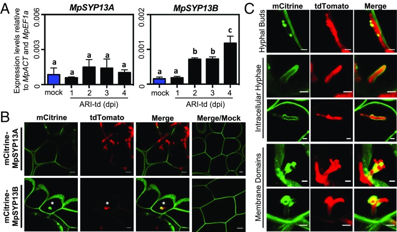
Fig. 4.
A colonization-induced host syntaxin accumulates at intracellular infection structures. (A) qRT-PCR analysis of MpSYP13A and MpSYP13B transcripts in mock-treated or P. palmivora-colonized (ARI-td) TAK1 plants from 1 to 4 dpi. Expression values are shown relative to internal MpACT and MpEF1a controls. Different letters signify statistically significant differences in transcript abundance (ANOVA, Tukey’s HSD, P < 0.05). (B) Confocal fluorescence microscopy demonstrating mCitrine-MpSYP13A/B localization in cells containing P. palmivora (ARI-td) intracellular infection structures at 3 dpi. Asterisks denote intracellular infection structures. (Scale bars, 10 μm.) (C) Patterns of mCitrine-MpSYP13B localization in P. palmivora-colonized (ARI-td) plants, including close-up images of the structure displayed in B. (Scale bars, 5 μm.) Experiments were performed three times, with similar results.

