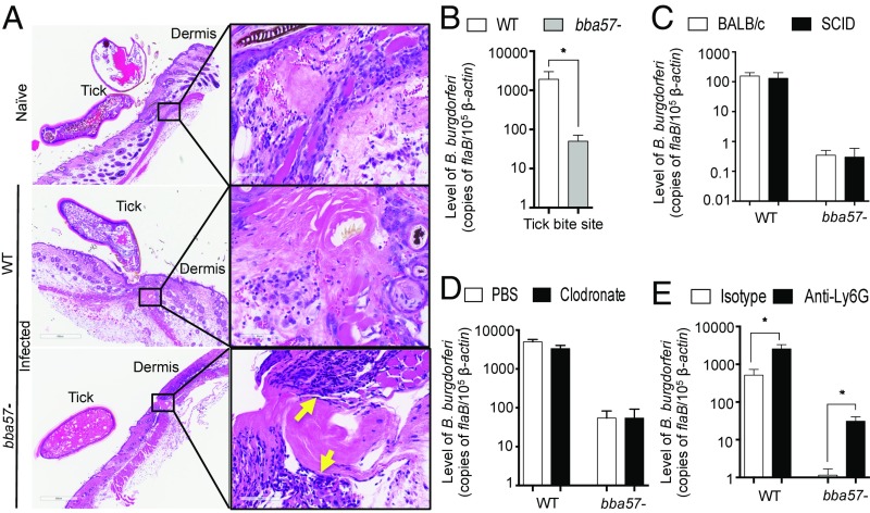
Fig. 4.
BBA57 protects B. burgdorferi at the dermal inoculum site against neutrophil-derived bactericidal responses. (A) Histology of murine skin at tick bite sites 5 d after placement of naive nymphal ticks or infested nymphs shows increased cellular infiltrates at the tick-bite site in those animals exposed to bba57− B. burgdorferi (yellow arrows). (B) B. burgdorferi burden at the tick bite site. The WT (white bars) or bba57− mutant B. burgdorferi (gray bars)-infected ticks were placed on naive mice. Five days later, the bite sites were collected and processed for bacterial burden analysis by RT-qPCR. (C) The levels of B. burgdorferi at the injection site in immunodeficient mice. Groups of BALB/c (white bars) or SCID mice (black bars) were injected with pathogens, and their burdens were detected at the injection site after 5 d by RT-qPCR. (D) Burden of B. burgdorferi at the injection site following macrophages depletion. Mice were treated with liposomes containing either PBS (white bars) or clodronate (black bars). Following cell depletion, mice were injected pathogens, and their burdens were detected after 5 d by RT-qPCR, as shown in C. (E) B. burgdorferi burden at the injection site after neutrophil depletion. Mice were treated with control (white bars) or anti-Ly6G (clone 1A8) antibodies (black bars) to deplete neutrophils. Following cell depletion, mice were injected pathogens, and their burdens were detected after 5 d by RT-qPCR, as shown in C. Bars represent the mean ± SEM of at least triplicates. Data in A–E are representative of at least six mice and two independent experiments. *P < 0.05.

