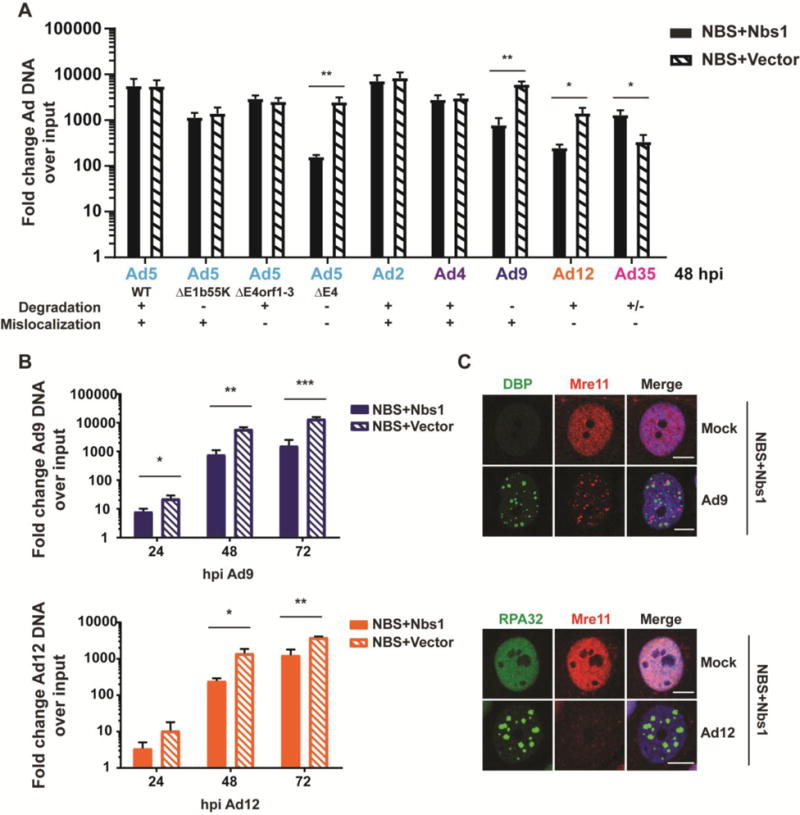
Figure 4. MRN impairs Ad9 and Ad12 replication.

(A) Hypomorphic Nbs1 cells complemented with wild-type Nbs1 (NBS+Nbs1) or empty vector (NBS+Vector) were infected to determine the effect of MRN on viral replication. Cells were harvested 48 hpi, and viral DNA accumulation was measured by quantitative PCR using primers specific for a conserved region of the viral genome. Values were normalized internally to tubulin and also to a 4-hour time point to control for input virus. Fold increase over input is shown, and error bars represent standard deviation from at least three biological replicates. Statistical significance was determined by a student’s T test (* = p < 0.05, ** = p < 0.01). (B) Viral DNA accumulation was measured in NBS+Vector and NBS+Nbs1 cells as in panel A over a time course of infection with Ad9 and Ad12. MRN impairs DNA accumulation at multiple time points of infection. Error bars represent standard deviation from at least three biological replicates. Statistical significance was determined by a student’s T test (* = p < 0.05, ** = p < 0.01, *** = p < 0.001). (C) Immunofluorescence of complemented NBS cells (NBS+Nbs1) 48 hpi confirms that Ad9 mislocalizes MRN and that Ad12 decreases MRN levels in these cells. Mre11 is shown in red. Viral DBP and cellular RPA32 (green) mark sites of viral DNA replication, and merged images include DAPI in blue. Scale bar = 10μm.
