
Figure 4. Apoptosis and caspase 3/7 activity in U-87 MG cells.
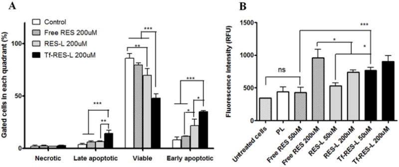
(A) U-87 MG cells were treated with RES formulations for 24 h before staining with AnV- Alexa Fluor 488® and PI and analyzed by flow cytometry. Untreated cells served as controls. The fluorescence of PI was recorded in the FL-3 channel and that of AnV was recorded in the FL-1 channel. Cells labeled with one or both the compounds were analyzed using a quadrant plot. (B) U-87 MG cells were treated with formulations for 24 h. After removal of the drug containing media, caspase reagent was added to the cells. The fluorescence was read after 4 h. The fluorescent product generated was proportional to the amount of caspase-3/7 cleavage activity of the sample. Data are represented as mean ± SD, from three separate studies. One-way ANOVA was used to compare between groups, p < 0.05 was considered significant (* p< 0.05, **p < 0.01, ***p< 0.001).