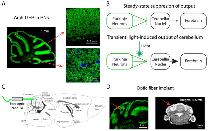
Figure 1. Light activation of Arch in Purkinje neurons drives cerebellar output during fMRI scans.
(A) Parasagittal cerebellar brain slice from an L7-Cre;RCL-Arch-GFP mouse showing selective Purkinje neuron expression of the Arch-GFP transgene (green). Arch-GFP expression is apparent in the membranes of Purkinje neuron soma and dendritic trees (right, top) as well as its axons (right, bottom) projecting to cerebellar nuclear neurons (blue, CamKIIa) (B) A schematic describing the use of inhibitory optogenetics to generate cerebellar output to the forebrain. At steady-state Arch expressing Purkinje neurons (green) tonically inhibit cerebellar nuclear neurons (black), thereby limiting cerebellar output to downstream sites like the forebrain. Synchronous pauses in discrete populations of Purkinje neurons during light activation of Arch disinhibits the cerebellar nucleus, thus driving cerebellar output. (C) Fiber optic cannulas were stereotaxically placed in the forelimb region of the cerebellar cortex at a 0° angle (horizontal) through a craniotomy in the caudal part of the occipital bone. (D) The implantation site is apparent in both exemplar histological section (left) and MRI structural scan (right) containing the cerebellum. The location of the implanted optic fiber (see red arrows) is visible as a dark area juxtaposed to the vermis near the medial simplex.

