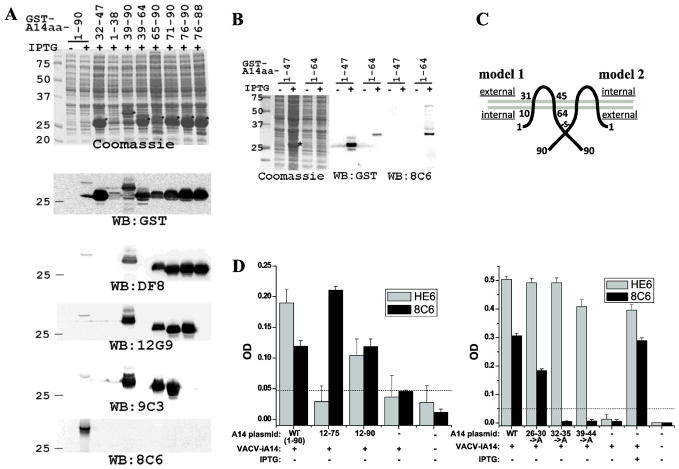
Figure 1. Mapping the epitopes of A14 mAbs. A and B).
Mapping the epitopes by Western blot of GST-A14 proteins. E. coli strains were either not induced (−) or induced with IPTG (+) to express GST fusion protein with the indicated A14 fragments. Proteins from the whole cell lysates were resolved by SDS-PAGE and analyzed by either Coomassie staining or by Western blot with the indicated antibodies. Prominent protein bands that are only present in induced samples are marked with *. Only Western blots with representative antibodies are shown. C). Predicted topology of A14 on MV with two possible orientations. The two grey lines represent the viral envelope, and the dark lines represent an A14 dimer. The amino acid residue numbers are indicated. The “-S-” denotes the disulfide bond via Cys71. The internal and external side of the virion are indicated in the two models. D). Further define the 8C6 epitope by ELISA of A14 mutants. 293T cells were transfected with plasmids encoding A14 alleles under the control of a VACV promoter and subsequently infected with an IPTG-inducible A14 mutant VACV (VACV-iA14) either in the absence of IPTG. The cell lysates were used to coat a microtiter plate, and ELISA was performed with either 8C6 or HE6. The A14 plasmids are named after the A14 residues expressed (12-75, 12-90) or A14 residues substituted with alanines (26–30->A, 32–35->A, 39–44->A).

