
Figure 1. Macrophages in the spiral ligament.
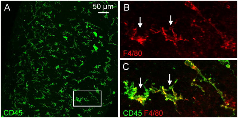
The images show macrophages in a whole-mount preparation of the spiral ligament collected from a young C57BL/6J mouse. The tissue was doubly stained with antibodies against CD45, a pan-leukocyte marker, and F4/80, a macrophage marker, using a method that has been previously described (Yang et al., 2015). A. Typical macrophages in the spiral ligament exhibit irregular shapes with large branches and short processes. B and C. Enlarged view of the macrophages in the area marked by the rectangle in A. B shows F4/80 immunoreactivity and C is the overlap of F4/80 and CD45 immunostaining of the same region shown in B.