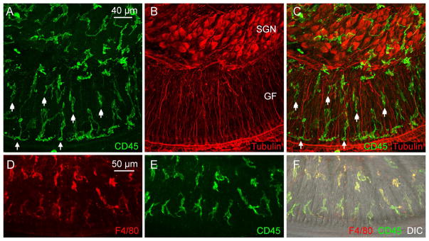
Figure 2. Identification of macrophages in the neural region inside the osseous spiral lamina and among ganglion cell bodies.
The images show macrophages in whole-mount preparations of cochleae in young C57BL/6J mice. The images were derived by projecting serial optical sections of confocal images covering only the depth of neural tissues in the osseous spiral lamina and ganglion cell regions. A. The image shows CD45 immunoreactivity in cochlear immune cells. B. Tubulin immunoreactivity is used to illustrate spiral ganglion neurons (marked by SGN) and their peripheral fibers (marked by GF, which stands for ganglion fiber). C. Merged view of A and B. Notice that macrophages are present in the regions of both ganglion cell bodies and their fibers. In the osseous spiral lamina, macrophages are arranged in two rows: one close to the edge of the osseous spiral lamina (shown by single-arrows) and the other close to ganglion neurons (marked by double-arrows). Many of these macrophages are oriented in parallel to the tunnel radial fibers of ganglion neurons. D, E, and F. Identity of macrophages is confirmed by the presence of F4/80 immunoreactivity. D shows the F4/80 immunoreactivity of immune cells. E. The same cells also display CD45 immunoreactivity. F. A merged view of A and B, as well as a differential interference contrast view (DIC) of the same region that illustrates the tissue orientation.

