
Figure 3. Identification of macrophages in the spiral limbus.
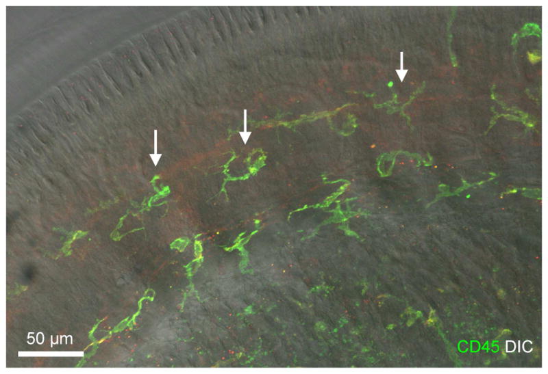
The image shows spiral limbus macrophages in a whole-mount preparation of a cochlea collected from a young C57BL/6J mouse. The image was derived by projecting serial optical sections of confocal image covering only the depth of the spiral limbus. The image is a superimposed image of CD45 immunostaining and DIC view of the same region. F4/80 immunoreactivity is weak and thus is not shown in the image. Macrophages identified in the spiral limbus region have a dendritic morphology with fine processes (marked by arrows).