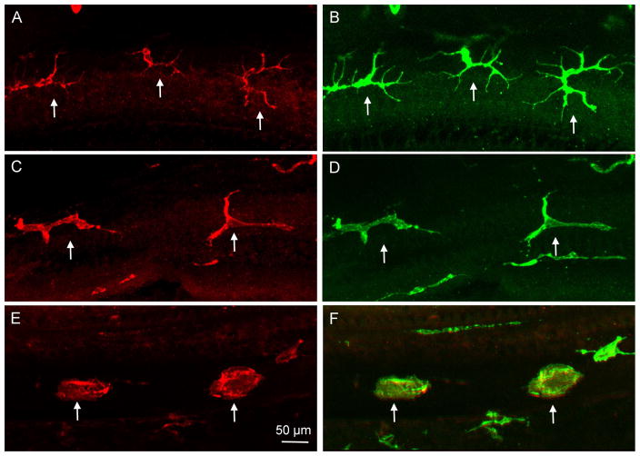
Figure 4. Distinct morphologies of basilar membrane macrophages along the apical-to-basal gradient of the cochlea.
The images show macrophages in a whole-mount preparation of the cochlea collected from a young C57BL/6J mouse. The tissues was stained for F4/80 and CD45. A. and B. Morphology of apical macrophages (approximately 0–30% distance from the apex). These cells display a dendritic shape with long thin projections (arrows). C and D. Morphology of macrophages in the middle region of the basilar membrane (approximately 30–70% distance from the apex). The long projections are shortened and the dendritic processes are reduced in number (arrows). E and F. Morphology of macrophages in the basal region of the basilar membrane (approximately 70–100% distance from the apex). Macrophages in this region display an amoeboid morphology without long projections or processes (arrows).

