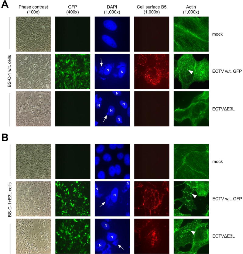
Figure 3. ECTVΔE3L infection of w.t. BS-C-1 cells resulted in an abortive replication cycle.
Normal BS-C-1 (A) or BS-C-1+E3L (B) cells were infected (MOI=10) with the indicated viruses. At 24 hrs post-infection, cells were examined for GFP expression, stained with DAPI to visualize virus factories (arrows) and cell nuclei (N), stained for B5 at the plasma membrane, or stained for actin. Examples of actin tails are indicated by the arrow heads. Representative images are depicted; each row does not necessarily show the same field of view.

