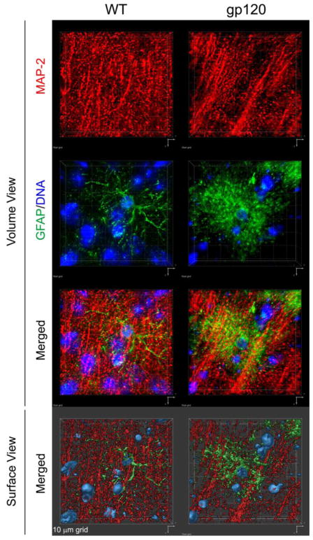
Fig. 1. Immunofluorescence staining of MAP-2-positive neuronal dendrites and GFAP-positive astrocytes in cerebral cortex of HIV gp120-transgenic and non-transgenic, wild type control mice.
Sagittal brain sections of 6 months-old gp120-transgenic and wild-type (WT) littermate controls were immune-stained for neuronal MAP-2 (red) and astrocytic GFAP (green). DNA (blue) was labeled with H33342 and is shown to indicate nuclei. Fluorescence-labeled sections were analyzed using a Zeiss Axiovert 100 M inverted microscope and Slidebook software (Intelligent Imaging Innovations, Denver, CO) to record Z-stacks and perform deconvolution and 3D reconstruction. The upper six panels show 3D volume views, the bottom two panel are 3D surface views. Representative areas of mid-frontal cortex, layer 3, are shown. Note the difference in the density of MAP-2 immunoreactive neuropil and astrocyte morphology between WT and gp120tg samples, and the dimensions of the 10 μm grid.

