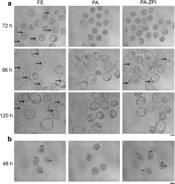
Fig. 4.

Different hatch patterns between fertilized and parthenogenetic embryos cultured in vitro. a Morphology of fertilized, normal and ZP-drilled parthenogenetic embryos cultured in vitro for 72, 96 and 120 h. Thick and black arrows indicated the place where embryos began to hatch. b Hypotonicity treatments with embryos cultured for 48 h. Thin and black arrows indicated the place where cytoplasm permeated out. FE, fertilized embryos cultured in 20% O2; PA, normal parthenogenetic embryos cultured in 20% O2; PA-ZPI, parthenogenetic embryos with a breach about 5 μm on ZP, drilled by a way like pronuclear injection with Piezo micromanipulator and cultured in 20% O2. Bar = 50 μm
