
FIGURE 2.
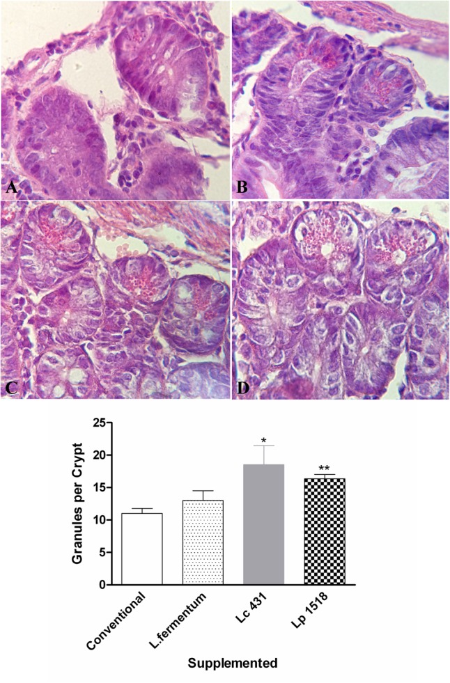
Micrographs of small intestine sections in the different animals’ models. Tissue sections of 42 days old mice fed with conventional diet (A), L. fermentum (B), L. casei CRL 431 (C), or L. paracasei CNCM-I 1518 (D) stained with hematoxylin and eosin. Magnification 1000×. Results (mean ± SEM) are representative of three independent experiments. The differences were calculated with respect to the control group that received conventional diet ∗p < 0.05, ∗∗p < 0.01.