
FIGURE 4.
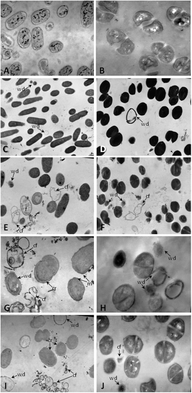
Transmission electron microscopy of pathogens undergoing to intestinal fluids of animals. Approximately 2 × 10⁸ CFU of (A,C,E,G,I) S. Typhimurium, (B,D,F,H,J) S. aureus were incubated for 1 h at 37°C in the presence of: (A,B) 10 mM sodium phosphate; the intestinal fluids of mice fed with: (C,D) conventional diet, (E,F) L. paracasei CNCM-I 1518, (G–I) L. casei CRL 431; and (J) Gentamicin 20 μg/ml. Magnification (C) 7500×; (A,D–F,I) 12.800×; (B,G,H,J) 22.800×. Cr, cytoplasmic retraction; f, electron-dense fibers; cf, cell fragmentation; wd, cell walls disruption; and m, membrane ruffling.