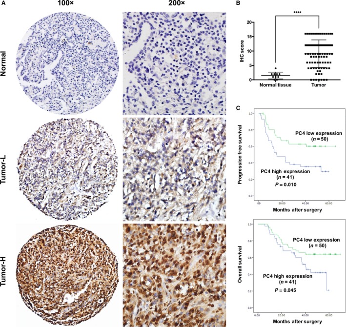
Figure 6.

Prognostic significance of PC4 expression in patients with NSCLC. (A) Normal: corresponding normal lung tissue showed low expression of PC4 (IHC score: 0). Tumor‐L: NSCLC tissue exhibited low expression of PC4 (IHC score: 6). Tumor‐H: NSCLC tissue exhibited high expression of PC4 (IHC score: 16). (B) Statistical analysis revealed significantly higher expression of PC4 in NSCLC tissues (**** P < 0.0001, Student's t‐test).(C) Kaplan–Meier plots showed progression‐free survival and overall survival curves of the 91 patients, according to PC4 expression levels in the primary tumor (P < 0.05, log‐rank test).
