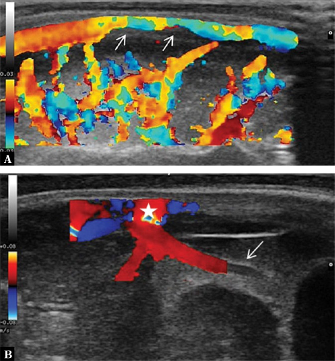
Fig. 3.
A. Color Doppler imaging shows increased flow in the superficial cerebral vessels. In the superior sagittal sinus flow constriction associated with the presence of a parietal thrombus (arrows) is seen. B. Imaging through the anterior fontanelle in the longitudinal plane. One of the perforator veins is dilated with a surrounding hyperechoic extra-axial echo. A vessel partly covered by the color Doppler gate with a visible ostium into the superior sagittal sinus (star) is seen

