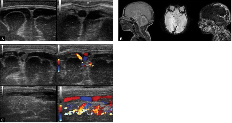
Fig. 8.
A twenty-six-day-old infant with diagnosed BM caused by Streptococcus agalactiae. In the course of the therapeutic process the diagnostic examination of the coagulation system was extended and concomitant protein S deficiency was diagnosed. Low-molecular-weight heparins were used for treatment. A. On the left, imaging through the anterior fontanelle reveals a normal image of the superior sagittal sinus. A susceptible to pressure, triangular lumen of the vessel is notable. On the right, a round section of the superior sagittal sinus which is not susceptible to delicate pressure of the transducer is seen (arrow). If no thrombus is visualized using standard viewing planes, such an image can suggest that thrombosis is present in a segment of the vessel which is not accessible to scanning. B. Parietal thrombus in the superior sagittal sinus visible in transverse and longitudinal planes in B-mode and color Doppler. C. MRI: visible signs of disseminated coagulation in the cerebral venous system including superficial vessels, deep vessels (straight sinus – arrow) and the confluence of sinuses

