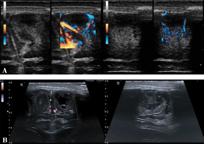
Fig. 10.
A. Acute phase of cortical infarction in the frontal gyri. A hypoechoic area without a visible flow in color Doppler with a surrounding hyperechoic area of reaction (on the left – transverse sections, on the right – longitudinal sections). B. Areas of subcortical leukomalacia. Color Doppler imaging shows no vascular flow in the visualized changes (on the left – transverse section, on the right – longitudinal section)

