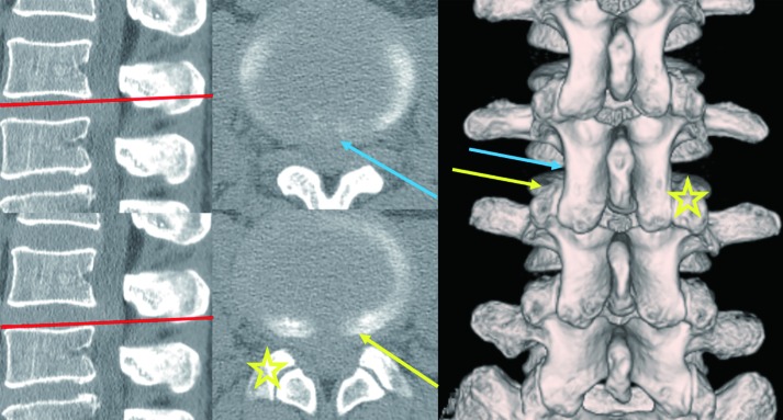
Figure 3.
Pre-operative CT finings of patients with accidental dural puncture during the initial operative step for discography. Sagittal, axial, and three-dimensionally reconstructed CT images. The red lines indicate the scanning positions; the blue arrow indicates the first needle trajectory for accidental dural puncture; the yellow arrow indicates the corrected needle trajectory for appropriate PELD via the TFA; the ☆ sign indicates the SAP of the contralateral side. Note that cranial needle puncture is easier to reach the medial site of the annulus fibrosus. CT, computed tomography; PELD, percutaneous endoscopic lumbar discectomy; TFA, transforaminal approach; SAP, superior articular process.

