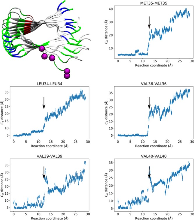
Figure 5.

Residues involved in the third peak of the PMF. Top left: Alpha carbons of Leu34, Met35, Val36, Val39, and Val40 represented as magenta-colored spheres. P0 and P1 are shown as ribbons colored by residue type (white for hydrophobic, green for hydrophilic, blue for positively charged, and red for negatively charged), while the rest of the peptides are shown in cartoon representation colored by residue type. Top right, bottom left and right: Distance between corresponding alpha carbons of P0 and P1 as a function of the reaction coordinate. Arrows highlight the transition, occurring at displacements consistent with the third peak.
