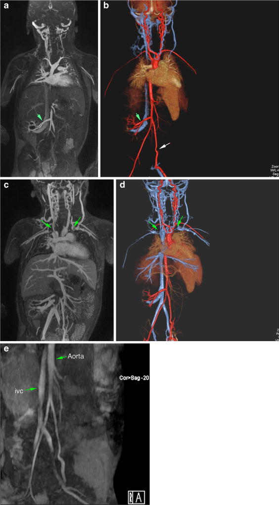
Fig. 1.
a–e Magnetic resonance (MR) angiogram with ferumoxytol in 3-year-old male post-renal transplant. Arterial phase-dominant (a, b) and venous phase-dominant (c, d) images show widely patent graft artery and vein and multiple thrombosed neck/chest veins (green arrows). For comparison (e) is a non-contrast, time-of-flight MR angiogram performed before the transplant, which highlights the superior clarity of the ferumoxytol studies

