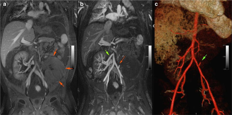
Fig. 3.
Magnetic resonance angiogram with ferumoxytol in an 8-year-old male post-second renal transplant. a Enlarged and edematous graft in the left lower quadrant (as denoted by green arrows). b Patent right renal artery to previous graft (green arrow) and a thrombosed left renal artery (red arrow) to the infarcted second graft. c Volume-rendered 3D image of the same patient, highlighting the thrombosed left renal graft artery (green arrow)

