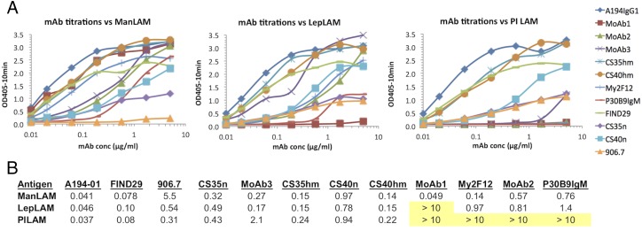
FIGURE 1.
(A) Binding titration of mAbs to purified LAMs isolated from M. tuberculosis (ManLAM), M. leprae (LepLAM), and M. smegmatis (PILAM). After coating the Ags (20 μg/ml) on ELISA plates, the plates were blocked by incubation with a solution of 1% BSA and titered against a panel of 10 mAbs reactive with LAM carbohydrate structures. Ab binding was detected with the appropriate AP-labeled secondary Ab. (B) Tabulation of the 50% maximum binding concentration (μg/ml) of each mAb for each of the Ags. Nonreactive combinations are highlighted in yellow.

