
Fig. 1.
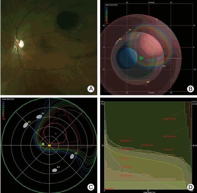
A case of proton beam therapy (PBT) planning in eyeball model of a patient with choroidal melanoma. (A) Fundoscopic finding suggested the tumor located near 2 disc diameter from macula. (B) Beams-eyed view. (C) Dose distribution of PBT in fundus view. (D) Dose volume histogram.