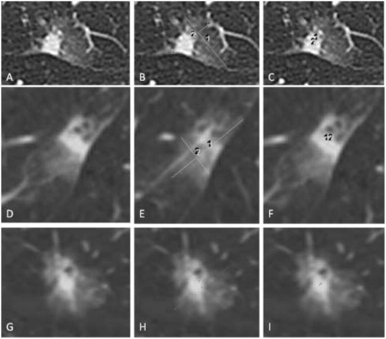
Figure 1.

Measuring lesions on CT. (A-C) Axial CT images of a subsolid lesion with (B) measurement of the whole lesion in maximum long axis and a perpendicular measurement and (C) measurement of the solid component in maximum long axis and a perpendicular measurement. (D-F) The lesion and measurements taken in the sagittal plane. (G-I) The lesion and measurements taken in the coronal plane.
