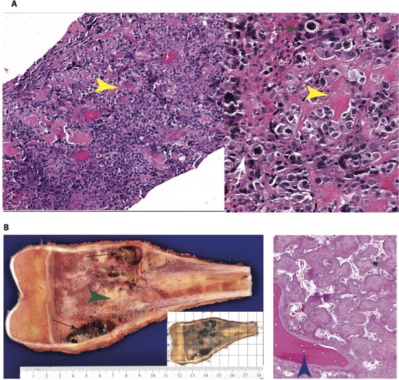
Figure 3.

Pathology of a high-grade intramedullary (conventional) osteosarcoma. (a) At low-(100x, left) and intermediate (200x, right) magnification, the diagnostic biopsy reveals a cellular malignancy with conspicuous malignant osteoid formation (yellow arrow). The cellular component is composed of pleomorphic cells with osteoblast-like morphology. Increased mitotic activity (white arrow) and individual cell necrosis (gray arrow) are evidence of rapid cell turnover. (b) Gross images of the resected specimen (left) reveal an expansile mass centered in the medullary cavity with areas of hemorrhage and cystic degeneration (black arrows) and neoplastic bone formation (green arrowhead). In routine processing of these specimens, a complete cross section is blocked out (grid, small inset) and submitted for histologic processing to assess treatment response. The histologic findings in the resection specimen (right) are typical of treated osteosarcoma. The cellularity is markedly diminished, with necrosis and neoplastic bone remaining. The marbled, irregular seams of osteoid have a paucicellular stroma and are distinct in appearance from the native, lamellar bone (blue arrowhead).
