Figure 5.

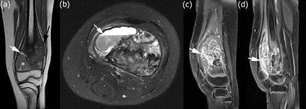
High-grade intramedullary (conventional) osteosarcoma of the left distal femur in a 12-year-old boy with distal femoral osteosarcoma after neoadjuvant chemotherapy. (a) Coronal T1-weighted MRI without fat saturation of the left knee. The lesion shows interval decrease in size of the soft tissue mass (black arrow). There are now new foci of increased T1 signal within the lesion consistent with hemorrhage (white arrow). An area of necrosis with hemorrhage and cystic change is seen in the medial metaphysis (white asterisk). (b) Axial T2-weighted MRI with fat saturation illustrating an area of necrosis with hemorrhage and cystic change with a fluid-fluid level (white arrow). There remains unchanged low T2 signal within the lesion corresponding to malignant bone formation (white asterisk). (c) Sagittal T1-weighted MRI of the left knee obtained after intravenous administration of contrast material reveals interval decrease in size of the soft tissue mass with decreased enhancing peritumoral edema and internal necrosis within the tumor after chemotherapy. (d) Sagittal T1-weighted MRI of the left knee obtained after intravenous administration of contrast material shows internal cystic change within the tumor.
