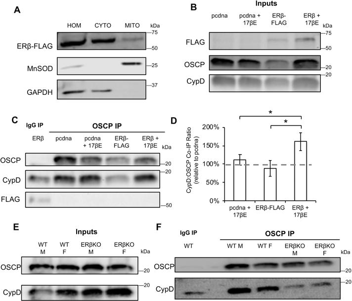
Figure 7. ERβKO decreases the interaction between OSCP and CypD in brain mitochondria.

A) Western blot of total homogenate (HOM), cytosolic (CYTO) and mitochondrial (MITO) fractions of cells transfected with ERβ-FLAG. Manganese SOD and GAPDH are used as mitochondrial and cytosolic markers, respectively. B) Western blot of FLAG, OSCP and CypD in enriched mitochondrial fractions from cells expressing empty vector (pcdna) or ERβ-FLAG, with and without 17βE treatment. C) Western blot of co-IP eluate from IgG IP or OSCP co-IP. D) Quantification of the ratio of CypD:OSCP in co-IP eluates. Data are expressed relative to the CypD: OSCP ratio in pcdna transfected cells. * p < 0.05 by one-way ANOVA with Bonferroni post-hoc analysis; n = 7 independent biological replicates. E) Western blot of OSCP and CypD in purified brain mitochondria from wild type (WT) and ERβKO males and females. F) Western blot of co-IP eluate from IgG IP or OSCP co-IP.
