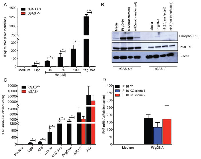
Figure 3. Pf gDNA induces IFNβ expression through the cGAS-STING pathway.
(A) cGAS+/+ and cGAS−/− THP-1 cells were transfected with 10, 50 or 100 μM of Hz for 6 h followed by detection of IFNβ mRNA by qRT-PCR. Pf gDNA was used as control. Data were analyzed for the difference in IFNβ mRNA induction in WT cells vs KO cells by paired Student’s t-test. (B) Phosphorylation of IRF3 was detected by SDS-PAGE followed by western-blot. Detection of total IRF3 and β-actin was used as loading control. (C) Transfection of AT-rich ODNs (AT5, AT5 3x and dsAT5 4x, 3 μM each) results in a cGAS dependent induction of type I IFN. Sendai virus (SeV) was used as control for cGAS-independent induction of IFNβ. (D) IFI16+/+ (EV-empty vector) and IFI16 KO (clones 1 and 2) THP-1 cells were transfected with 10 μg/ml of Pf gDNA for 6 h, then IFNβ mRNA was measured by qRT-PCR. Data were analyzed for the difference in IFNβ mRNA induction in WT cells vs KO cells by Wilcoxon rank test. Data are presented as mean ± SD and are representative of 3 independent experiments. * indicates a p value of < 0.05, ** indicates a p value of < 0.001.

