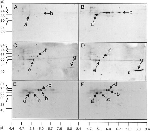
Figure 5.
Immunodetection from two-dimensional gels of vacuolar and cell wall invertase in mature leaf (A–D) and primary root (E and F) from well-watered (left column: A, C, and E) or water-stressed plants (right column: B, D, and F) for 7 d . Homologous groups of spots were designated a, b, c, and d for vacuolar invertase antibodies and e, f, and g for cell wall invertase antibodies. Antiserums raised against an IVR2 oligopeptide (A and B for mature leaf; E and F for primary root) and a cell wall invertase peptide (C and D for mature leaf) were used for invertase immunodetection from crude protein extracts (50 μg) in mature leaf and root. All sampling was done at 9 am. Comparison among loaded protein quantities was carried out from Coomassie Blue gel staining (data not shown). To measure pI, four gels were cut into 15 fragments and four gel fragments were incubated together in 1 mL of distilled water overnight; pH was measured from these eluted solutions.

