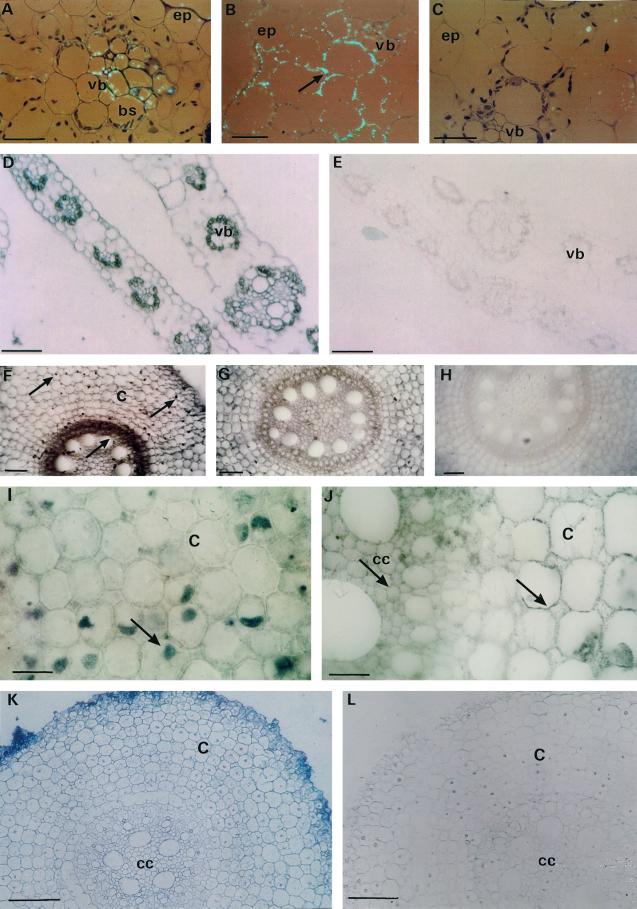
Figure 9.
In situ immunolocalization of invertase protein (A–C and F–J) and in situ hybridization of Ivr2 mRNA transcripts (D–E and K–L) in mature leaves (A–E) and roots (F–L) sampled on d 7 from water-stressed plants. Epipolarization optics of water-stressed mature leaf section exposed to A, Vacuolar invertase antibodies showing intracellular labeling in the vascular bundle; B, cell wall invertase antibodies showing cell wall labeling (arrow); and C, nonimmune serum. D, leaf section of water-stressed mature leaf hybridized to Ivr2 probe, in antisense orientation; E, leaf hybridized to Ivr2 probe in sense orientation. F and I, Root section of water-stressed plants incubated with vacuolar invertase antibodies showing intracellular localization in cortex and central cylinder (arrows); G and J, water-stressed roots exposed to anti-cell wall invertase serum showing immunopositive cell walls in all root tissue (arrows in J); H, root section incubated with nonimmune serum yielded no labeling; K, root section of water-stressed plants hybridized to Ivr2 antisense probe; L, root hybridized to sense probe. c, Cortex cells; cc, central cylinder; ep, epidermis; bs, bundle sheath cells; vb, vascular bundle cells. Bars in A through C, I, and J, 25 μm; D through H, K, and L, 100 μm.

