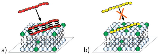
Figure 1.
Schematic diagram for Arg9 (red) and Lys9 (yellow) interacting with a supported lipid bilayer containing negatively charged phosphatidylglycerol lipids (dark green). (a) Arg9 displays much less pronounced anticooperative binding behavior, which is consistent with favorable short-range interactions (dashed lines) between the guanidinium moieties on the peptide, whereas (b) Lys9 experiences strong anticooperative binding effects.

