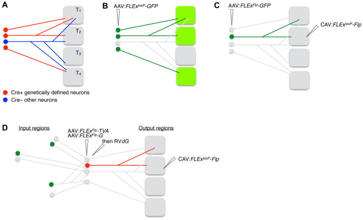
Figure 4. Cell Type-specific Axonal Projection and Collateralization Patterns.
(A) Ground truth of the axonal projection patterns (right) of neuronal populations at a specific brain region (left). T1–T4: target regions 1–4.
(B) Injecting AAV expressing Cre-dependent (FLExloxP) GFP into Cre transgenic mice can determine the axonal projection patterns of all Cre+ neurons at the AAV injection site. In this and all subsequent panels, unlabeled neurons are in grey, and their axons are omitted.
(C) Injecting retrograde transducing canine adenovirus 2 (CAV) expressing Cre-dependent Flp at one of the targets, along with injecting AAV expressing Flp-dependent (FLExFRT) GFP at the cell body region of Cre transgenic mice, can determine the complete axon collateralization patterns of Cre+ neurons that project to a specific target region.
(D) Replacing Flp-dependent GFP in (C) with Flp-dependent TVA and rabies glycoprotein (G), followed by RVdG at the cell body region enables trans-synaptic tracing of inputs (green) to Cre+ neurons that project to a specific output region, a strategy named cell-type-specific tracing the relationship between input and output (cTRIO).

