
Figure 5. Cell Type-specific Imaging.
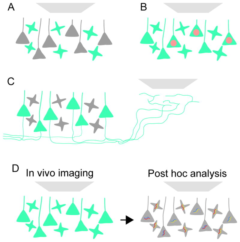
(A) Genetically encoded indicator of neural function is targeted to specific cell types (e.g. a local interneuron) for imaging.
(B) The indicator is expressed in all neurons for imaging. One cell type (e.g. a subtype of pyramidal neuron) in addition is identified with a fluorescent marker (in this example a red fluorescent protein targeted to the nucleus).
(C) The indicator is expressed in all projection neurons, but imaging is performed in the projection zone of one of the projection neuron types.
(D) The indicator is expressed in all neurons for imaging. Cell types are identified post hoc using molecular analysis (e.g. multiplexed fluorescent in situ hybridization).