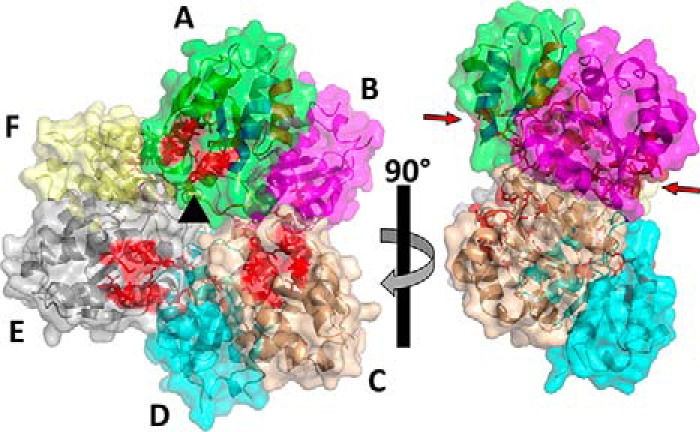
Figure 3.

Structure of the B. subtilis MgsA hexamer. The hexamer is composed of three dimers of MgsA, which arrange in the hexamer with three active sites (active-site residues indicated in red) arranged by 120° rotations (indicated by the black triangle in the center) on either side of the hexamer. The individual molecules are indicated in different colors and labeled A–F (top right panel). Bottom right panel, side view of the hexamer. The arrows indicate the active sites of molecules A and B.
