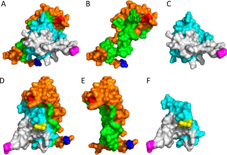
Figure 7.
Homology modeling of the CCL8-P672 complex. This is a 3D structural model of P672 (orange and green) and CCL8 (gray and cyan) modeled as a 1:1 complex using the EVA1-CCL3 structure (Protein Data Bank ID 3FPU) as template and MODELLER with default parameters after alignment with the MUSCLE algorithm. The residues forming the binding surfaces predicted using Arpeggio (23) are indicated in green (P672) and cyan (CCL8). The N terminus residue of P672 (Val-1) is indicated in blue and the C terminus residue (Trp-104) in red. The N and C termini of CCL8 are indicated in yellow and magenta, respectively. A and D, P672-CCL8 complex shown in two orientations. B and E, P672-alone shown in two orientations. The position of residue Asn-34, which is in close proximity to the C-terminal Trp-104, is indicated. C and F, CCL8 alone shown in two orientations.

