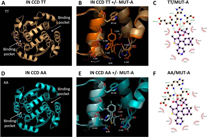
Figure 4.
Structures of IN CCD TT and AA ± MUT-A. A, global view of the IN CCD TT structure. B, superposition of the IN CCD TT ± MUT-A structures, close view of the binding pocket (gold = + ligand; light gold = − ligand). Large displacement of Thr-124 and Glu-170 between + and − MUT-A. C, 2D view of MUT-A interactions with IN CCD TT. D, global view of the IN-CCD AA structure. E, superposition of the IN-CCD AA ± MUT-A structures, close view of the binding pocket (light blue = + ligand, blue = − ligand). F, 2D view of MUT-A interactions with IN-CCD AA.

