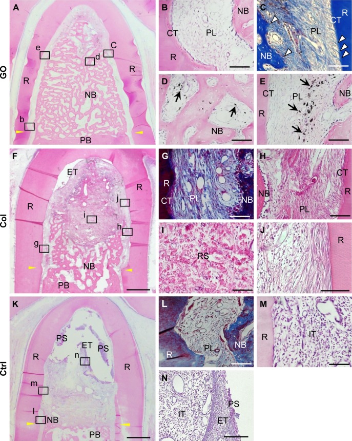
Figure 5.
Histological findings after implantation of GO scaffold into dog class II furcation defects.
Notes: (A–E) GO scaffold. High magnification (B–E) of the framed areas (b–e) in (A). Periodontal ligament-like tissue, cementum-like tissue, and alveolar bone with the insertion of Sharpey’s fibers (white arrowheads) was demonstrated. Black spots (black arrows) were found in the newly formed bone marrow and periodontal ligament-like tissue. (F–J) Collagen scaffold. High magnification (G–J) of the framed areas (g–j) in (F). (K–N) No implantation. High magnification (L–N) of the framed areas (l–n) in (K). Staining with H&E (A, B, D–F, H–K, M, N) and Masson’s trichrome (C, G, L). Scale bars represent 1 mm (A, F, K) and 100 μm (B–E, G–J, L–N). Yellow arrowheads (A, F, K) indicate the apical bottom of the periodontal defect.
Abbreviations: Col, collagen; CT, cementum-like tissue; Ctrl, control; ET, epithelial tissue; GO, graphene oxide; IT, inflammatory tissue; NB, new bone; PB, preexisting bone; PL, periodontal ligament-like tissue; PS, plaque-like structure; R, root; RS, residual scaffold.

