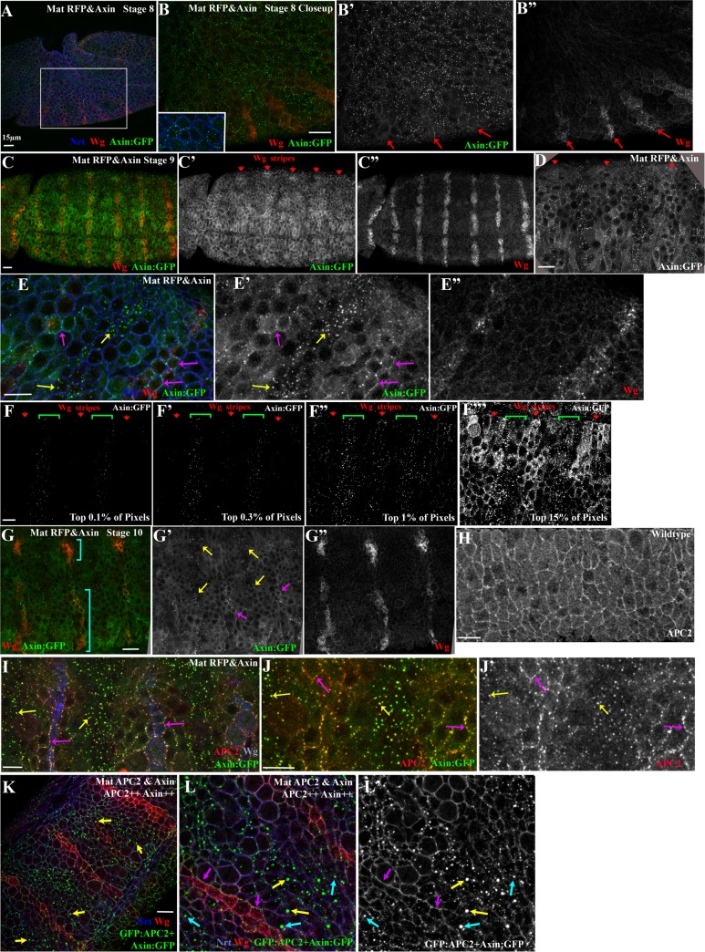
Fig 8. Axin assembles into cytoplasmic multiprotein destruction complexes together with APC2, and Wg signaling leads to their membrane-recruitment and elevates levels of cytoplasmic Axin.
(A,B) Fixed stage 8 Mat RFP&Axin embryo. Axin:GFP accumulates in puncta in all cells. Anterior to the left. Inset = close-up of B. Red arrows = initiation of Wg stripes. (C-E) Fixed stage 9 Mat RFP&Axin embryos. Anterior to the left. Red arrows = Wg expressing cells. (D,E) Close-ups of embryo in C showing Axin:GFP localization change in response to Wg. Yellow arrows = cytoplasmic puncta. Magenta arrows = membrane-associated Axin:GFP puncta. (F-F”‘) Image thresholding to determine the relative brightness of different pools of Axin:GFP. (F,F’) The brightest Axin:GFP pixels are in the cytoplasmic puncta in the interstripe cells (brackets). (F”) The next brightest pixels are in membrane–associated puncta in the Wg stripe cells (arrows). (F’”) Diffuse cytoplasmic staining is higher in Wg-stripe cells (arrows) than in interstripes (brackets). (G) Fixed stage 10 embryo. As the Wg stripe separates into medial and lateral domains (brackets), Axin:GFP continues to exhibit differential localization near or distant from Wg-expressing cells. Anterior to the left. Yellow arrows = cytoplasmic puncta. Magenta arrows = membrane-associated Axin:GFP puncta. (H) Localization of endogenous APC2 in a wildtype embryo. (I,J) Expression of Axin:GFP leads to recruitment of endogenous APC2 into both membrane-associated puncta in Wg-ON cells (magenta arrows) and into cytoplasmic puncta in Wg-OFF cells (yellow arrows). (K,L) Mat APC2&Axin. L = closeup. Presumptive APC2++ Axin++ embryo. Simultaneously highly elevating levels of both APC2 and Axin enhances resistance of the destruction complex to be turned off by Wg signaling. Yellow arrow = very bright cytoplasmic puncta. Cyan arrows = bright puncta found near Wg-positive cells. Magenta = membrane-associated puncta in Wg expressing cells. Scale bars = 15μm.

