
Fig. 2.
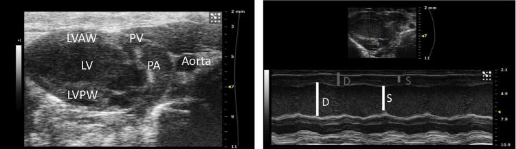
(Left) Parasternal long axis view of the left ventricle in B mode. LVAW = Left ventricle anterior wall, PV = pulmonary valve, PA = pulmonary artery, LV = left ventricle, LVPW = left ventricle posterior wall. (Right) Parasternal long axis view of the left ventricle in M mode. In the thumbnail above, the yellow dashed line represents the indicator line positioned at the widest portion of the LV chamber at the level of the aorta. In the M mode view below, measurements of the LV internal diameter (LVID) can be performed at diastole (D) and at systole (S), represented by the white lines. The LV anterior (LVAW) and posterior wall (LVPW) dimensions can also be calculated at diastole and systole, represented by the gray lines. (Color version of figure is available online.)