
Fig. 5.
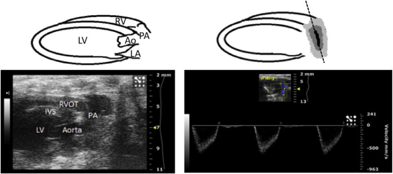
(Left) Modified parasternal long axis view demonstrating the right ventricular outflow tract and pulmonary artery in B mode of the ultrasound. (Right) Same view in color Doppler mode, used to assess flow through the pulmonary artery. The PW line is seen along the pulmonary artery on the right, positioned parallel to the direction of blood flow in the vessel, and can also be positioned to assess flow across the pulmonary valve and right ventricular outflow tract. RV = right ventricle; RVOT = right ventricular outflow tract; PA = pulmonary artery; LV = left ventricle; IVS = interventricular septum; Ao = aorta; and LA = left atrium. (Color version of figure is available online.)