
Figure 2.
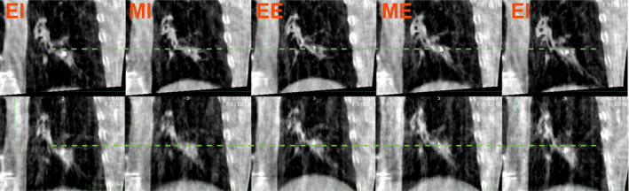
Example 4DCBCT phase images from the dataset, subject P105. Top row: First fraction 4DCBCT. Bottom row: 4DCBCT during 32nd fraction. The dotted line shows the superior tumor border at end of inhalation. The amplitude of motion is higher in the later 4DCBCT, as evidenced by a more superior position and end of exhalation. Left to right, approximate breathing phase: EI ‐ End inhalation phase, MI ‐ Mid inhalation phase, EE ‐ End exhalation phase, ME ‐ Mid exhalation phase, EI. These correspond to the 0%, 20%, 40%, 60%, and 80% phases, respectively.