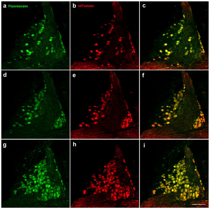
FIGURE 2.
All Phox2b-tdTomato-positive neurons (red) were retrogradely labeled in the geniculate ganglion following placement of tracer (green) on chorda tympani and greater superficial petrosal nerves. Retrogradely labeled neurons are shown in the ventral (a), middle (d), and dorsal (g) geniculate ganglion in single 1 μm optical slices taken from a whole mount geniculate ganglion. Phox2b-tdTomato-positive neurons (b, e, h). Double-labeled neurons (c, f, i). Scale bar in (i) =100 μm and applies to all images

