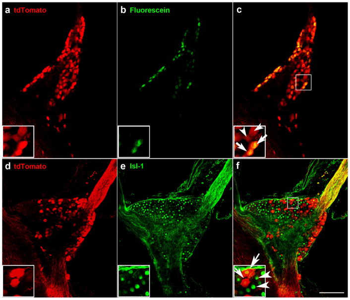
FIGURE 3.
Some but not all Phox2b-tdTomato-positive neurons in the geniculate ganglion were retrogradely labeled following placement of fluorescein on the chorda tympani nerve (a–c). A single optical slice through the geniculate ganglion whole mount shows Phox2b-tdTomato-positive neurons (a), retrogradely labeled neurons from the chorda tympani nerve (b), and the overlap between Phox2b-tdTomato and retrograde labeling was observed in some geniculate ganglion neurons (arrows in inset of c). There were also some Phox2b-tdTomato neurons without retrograde labeling (arrowheads). Expression of Phox2b-tdTomato was observed in some but not all geniculate ganglion neurons (d–f). Single optical slices through the geniculate ganglion stained for tdTomato (d), and the neuronal marker, Isl-1 (e). In the inset of (f), arrows indicate overlap between Isl-1 and tdTomato, and arrowheads indicate Isl-1-only labeled neurons. Scale bar in (c) =100 μm and applies to all images

