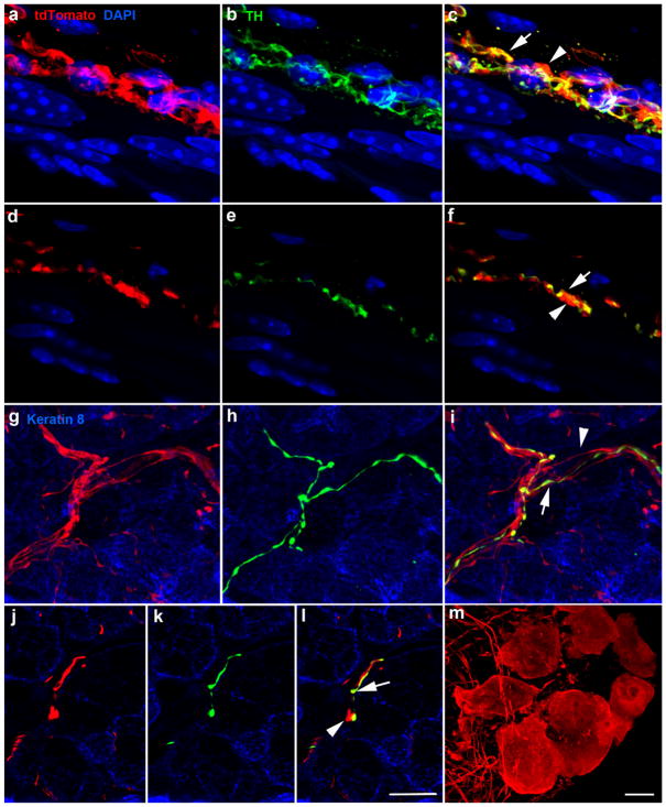
FIGURE 7.
Image z-stack (70 μm) of blood vessel innervated by tdTomato-positive fibers (red, a), TH-positive fibers (green, b), and overlay (c). TH-negative/tdTomato-positive and TH-positive/tdTomato-positive fibers run alongside each other. A single optical section from the z-stack illustrates individual TH-negative/tdTomato-positive fibers (arrowhead) and TH-positive/tdTomato-positive (arrow) (d–f). Innervation of Von Ebner’s glands was similar to that of blood vessels in that TH-negative/tdTomato–positive fibers (arrowhead) ran closely alongside TH-positive/tdTomato double-labeled fibers (arrow) (g, i). Single optical slices of von Ebner’s gland illustrating a single TH-negative/tdTomato-positive fiber (arrowhead) and TH-positive/tdTomato-positive fibers (arrow) (j–l). Cell bodies of the lingual parasympathetic ganglia were also tdTomato-positive (m). Scale bar in (l) =10 μm and applies to (a–l). The scale bar in (m) =10 μm

