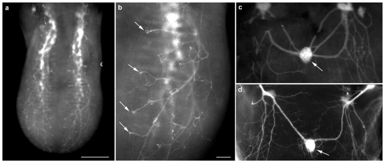
FIGURE 8.
Whole tongue from an embryonic day 14.5 Phox2b-Cre:tdTomato mouse. Without any additional labeling, fiber bundles could be seen entering the base of the tongue and branching near the surface (a). Fibers could be seen projecting to specific locations on the tongue surface where fungiform papillae were located (arrows) (b). tdTomato-positive innervation was also seen innervating the circumvallate papilla (arrow) in the back of the same tongue (c) and innervation has a similar pattern as seen in fixed DiI-labeled tissue (d). Scale bar: (a) =200 μm, (b) =250 μm (applies to c and d)

