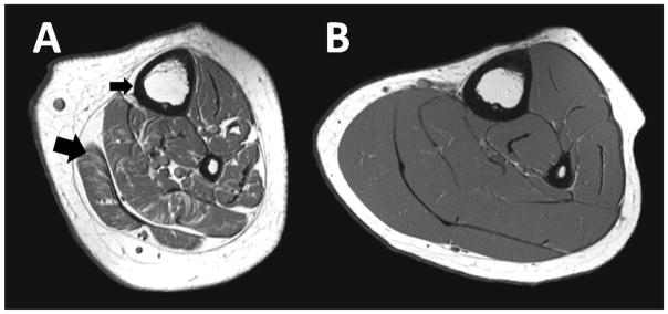
Figure 5.
Raw T1-weighted magnetic resonance images from the midtibia demonstrate the marked deficit in bone architecture and muscle volume and the high infiltration of fat within and around the musculature in an ambulatory boy with mild CP (A) compared to a typically developing boy with the same tibia length (B). In the image of the child with CP (A), the small black arrow highlights the thin cortical shell and the large arrow highlights the fat infiltration of muscle.

