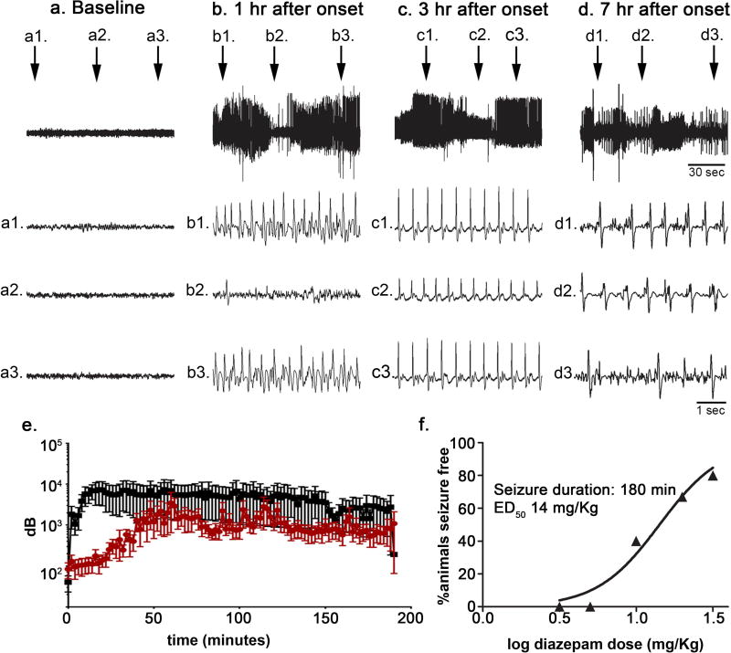
Figure 2. Kainic acid-induced SE of 180 min duration was also successfully treated with diazepam.
A: Hippocampal EEG recording obtained from a single animal prior to (a) and at various time points (b, c, d) following the onset of kainic acid-induced SE. Top trace, 150-sec epoch. Second trace (a1, b1, c1, d1), third trace (a2, b2, c2, d2), and fourth trace (a3, b3, c3, d3), 5-sec epoch at time indicated in the top trace. B, total power spectra during SE induced using lithium-pilocarpine (black) or kainic acid (red). C, Relationship between diazepam dose and percentage (n = 5 animals per dose per seizure duration) of animals seizure free at a time point 30 minutes following diazepam administration without seizure recurrence for an additional 90 minutes when diazepam was administered 180 minutes after kainic acid-induced seizure onset. The data were fitted to a sigmoidal dose-response curve with the maximum fixed to 100% and the minimum to 0. The median effective dose (ED50) value was derived from the best-fit equation.

